• Published on

    IMR

    IAPMD NEWS • ACHIEVEMENT

    Congratulations! IMR's Success at the IAPMD 11th Annual Scientific Meeting 2026

    August 2026 • Kuala Terengganu, Malaysia
    The Institute for Medical Research (IMR) extends its heartiest congratulations to Dr. Ainur Yusniza Yusof from the Autoimmune Unit, Allergy & Immunology Research Centre (AIRC) for winning the Bronze Award for Best Poster Presentation at the IAPMD 11th Annual Scientific Meeting 2026.
    Best Poster Presentation • Bronze Award
    Serological Characterization and Clinical Significance of Onconeuronal Autoantibodies in Suspected Paraneoplastic Syndrome
    Awardee: Dr. Ainur Yusniza Yusof
    Unit/Centre: Autoimmune Unit, Allergy & Immunology Research Centre (AIRC), IMR
    Category: Original Article
    Award: Best Poster Presentation Award – Bronze Award

    The scientific meeting took place on 3–4 August 2026 at the Raia Hotel & Convention Centre, Kuala Terengganu, Terengganu.

    The award recognises the scientific contribution of Dr. Ainur Yusniza Yusof and her research team in investigating the serological characteristics and clinical significance of onconeuronal autoantibodies in patients with suspected paraneoplastic syndrome.

    This achievement highlights the valuable contribution of medical research and pathology-related scientific work in advancing diagnostic understanding and improving patient care.

    “Excellence in medical research is the catalyst for elevating the quality of national healthcare.”
    Kudos and warmest congratulations to Dr. Ainur Yusniza Yusof!

    May this achievement continue to inspire researchers across the Institute for Medical Research and the Ministry of Health Malaysia to excel on scientific platforms locally and internationally.
    ```
  • Published on

    CPATH

    International Academy of Pathology — Malaysian Division

    IAPMD Achievement

    Lifetime Achievement Award
    August 31, 2025

    Congratulations to Prof Emerita Dr Nor Hayati Othman

    Our sincere congratulations to our President, Prof Emerita Dr Nor Hayati Othman, for being the recipient of the College of Pathologists, Academy of Medicine Malaysia Lifetime Achievement Award.

    The award recognises her outstanding contribution to pathology and the medical profession, as well as her dedication to the advancement of pathology in Malaysia.

    A remarkable achievement and a proud moment for IAPMD.
    ```
  • Published on

    Sunway Medical Centre

    International Academy of Pathology — Malaysian Division

    10th IAPMD Scientific Meeting

    October 2, 2025

    10th IAPMD Scientific Meeting at Sunway Medical Centre

    We were proud to be the venue sponsor of the 10th Scientific Meeting of the International Academy of Pathology – Malaysian Division (IAPMD), held on 1–2 October 2025 at the SunMed Convention Centre.

    The meeting brought together pathologists and healthcare professionals for scientific exchange, professional networking and continued learning.

    The event provided an excellent opportunity for members of the pathology community to share knowledge and strengthen professional connections.
    ```
  • Published on
  • Published on

    Case 9 : Mesonephric hyperplasia

    Picture

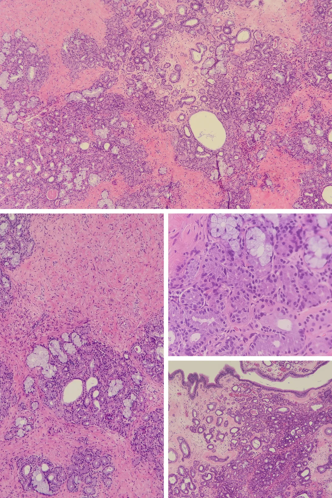
    A 46-year-old lady was diagnosed with adenocarcinoma in situ on pap smear and she underwent cervical cone biopsy. One representative section.

    ​Educational notes: 1. Section from the cervical cone biopsy shows mesonephric hyperplasia characterized by lobules of glands in the deeper part of the cervical wall. These glands are composed of a single layer of cuboidal epithelium without cytological atypia. The covering squamous epithelium and endocervical glands are devoid of dysplasia. 2. Presence of lobules of glands in the deeper part of the cervical wall stimulates infiltrative adenocarcinoma. However, entities such as mesonephric remnants and mesonephric hyperplasia need to be considered. Mesonephric remnants typically involve the deep layer of anterolateral wall of cervix along the path of the mesonephric duct. They may appear infiltrative and extend close to the luminal surface and intermingle with endocervical glands. In contrast, mesonephric hyperplasia has larger and more irregularly distributed glands as compared to mesonephric remnants. In the lobular type, the glands have exaggerated, clustered, lobular arrangement separated by variable amount of stroma. In the diffuse type, mesonephric hyperplasia displays predominantly non-clustered, extensive, and more diffuse proliferation. The least common duct type is characterized by hyperplastic epithelium with papillary tufting, and it may show clefted contours lacking intraluminal secretions. 
    3. Mesonephric glands are characteristically lined by a layer of simple flat or low cuboidal, non-ciliated cells with distinct basement membranes. They contain PAS-positive, diastase resistant material. Differing from adenocarcinoma or adenocarcinoma in situ, mesonephric glands lack nuclear atypia. Adenocarcinoma in situ is consistently characterized by nuclear enlargement, coarse chromatin, small single or multiple nucleoli, increased mitotic activity and variable nuclear stratification. An invasive adenocarcinoma shows complex architecture forming labyrinth or maze-like arrangement, and cribriform pattern with single gland profile. Although presence of stromal desmoplasia favors malignancy, distinction from a range of benign conditions showing stromal fibrosis, oedema and dense inflammation is critical.

    ​Reference 1. Zaino, R. Glandular Lesions of the Uterine Cervix. Mod Pathol 13, 261–274 (2000).