• Published on

    Case 8 : Cerebral toxoplasmosis

    Picture
    ​Clinical History: A 33-year-old male with underlying retroviral disease presented with altered behavior. Biopsy of the brain lesion. One representative section.
    Educational notes:
    1. The brain tissue fragments show necrotizing cerebritis with large areas of necrosis containing eosinophilic cellular debris. There are foci of mixed acute and chronic inflammatory infiltrates and perivascular inflammatory infiltrates. Extracellular microorganisms and rare pseudocysts are observed, morphologically consistent with free tachyzoites and bradyzoites of Toxoplasma gondii.
    2. Most common differential diagnoses for intracranial lesions of AIDS patients are primary central nervous system lymphoma, progressive multifocal leukoencephalopathy, cerebral toxoplasmosis and HIV encephalitis. In this brain tissue biopsy, microorganisms with the size less than a lymphocyte are identified. Histoplasma capsulatum and Toxoplasma gondii are the main differentials. Morphologically, the former is distinguished from the latter by presence of fungal cell wall in the form of halo in H&E stain. This could be highlighted by GMS and PAS special stains. Toxoplasma gondii may be difficult to be recognized due to resemblance to cellular debris. Immunohistochemistry against Toxoplasma gondii is helpful.

    ​Reference 1. Lee, Ashley M., et al. "Safety and diagnostic value of brain biopsy in HIV patients: a case series and metaanalysis of 1209 patients." Journal of Neurology, Neurosurgery & Psychiatry 87.7 (2016): 722-733. 2. Kradin, Richard L. Diagnostic Pathology of Infectious Disease. Elsevier Health Sciences, 2017.
  • Published on
  • Published on

    Case 7-Giant cell glioblastoma, WHO grade IV

    Picture

    32 y.o., female, multiple hyperdense lesions on the right parietal region. One representative section.Immunohistochemistry shows the malignant cells are positive for GFAP and negative for pan-cytokeratin and HMB-45.

    Educational notes:
    1. Section shows tumor tissue fragments composed of bizarre malignant cells with angulated hyperchromatic nuclei and abundant cytoplasm. Numerous multinucleated tumor giant cells are admixed. Mitoses are easily observed. The adjacent glial tissue is focally infiltrated by malignant
    cells. Immunohistochemistry shows the malignant cells are positive for GFAP and negative for pan-cytokeratin and HMB-45. Histological findings are consistent with giant cell glioblastoma, WHO grade IV. (Note: Without IDH evaluation, a diagnosis of glioblastoma, NOS, is more appropriate, vide infra)
    2. The 2016 World Health Organization Classification of Tumors of the Central Nervous System employs integrated phenotypic and genotypic parameters for tumor classification. Glioblastomas are classified into (1) glioblastoma, IDH-wildtype (about 90 %), which corresponds to the clinically defined primary or de novo glioblastoma in older patients; (2) glioblastoma, IDH-mutant (about 10%), which corresponds to secondary glioblastoma with a history of prior lower grade diffuse glioma in younger patients, and (3) glioblastoma, NOS, a diagnosis for those tumors that IDH
    evaluation cannot be performed.
    3. Along with gliosarcoma and epithelioid glioblastoma, giant cell glioblastoma is a variant under the umbrella of glioblastoma, IDH-wildtype. Histologically, it is characterized by bizarre, multinucleated giant cells and an occasionally abundant reticulin network. Palisading and large
    ischaemic necroses are observed. Atypical mitoses are frequent. Microvascular proliferation is not common. Giant cell glioblastoma has a somewhat better prognosis than ordinary glioblastoma.
    Reference:
    1. World Health Organization. (2016). WHO Classification of Tumours of the Central Nervous System Revised 4th Edition. David N. Louis, (Ed.). Internat. Agency for Research on Cancer.
  • Published on

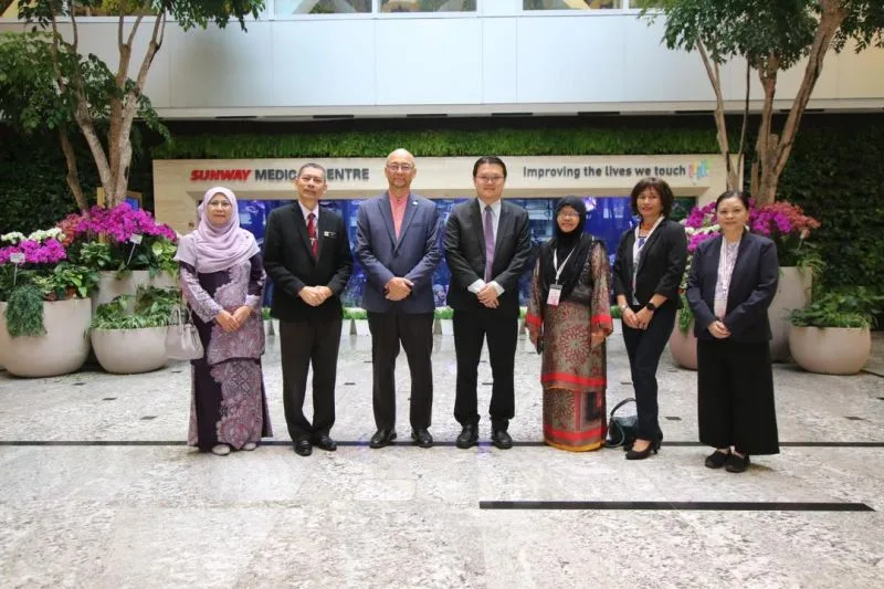
    Sunway Medical Centre

    International Academy of Pathology — Malaysian Division

    10th IAPMD Scientific Meeting

    October 2, 2025

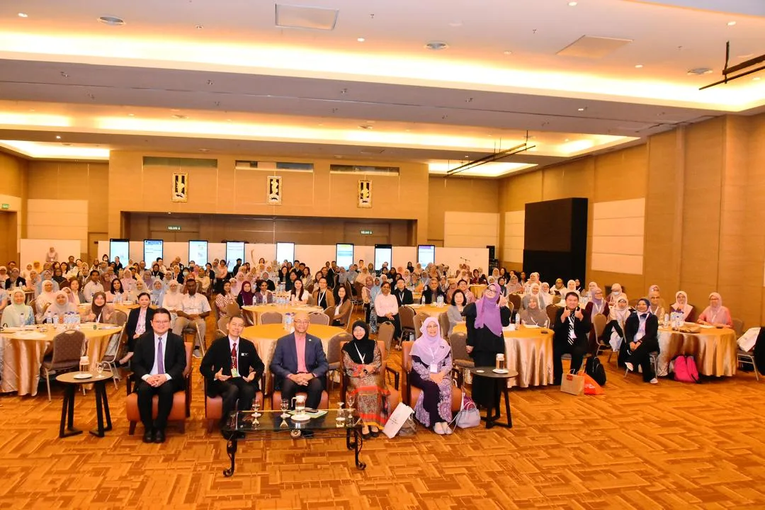
    10th IAPMD Scientific Meeting at Sunway Medical Centre

    We were proud to be the venue sponsor of the 10th Scientific Meeting of the International Academy of Pathology – Malaysian Division (IAPMD), held on 1–2 October 2025 at the SunMed Convention Centre.

    The meeting brought together pathologists and healthcare professionals for scientific exchange, professional networking and continued learning.

    The event provided an excellent opportunity for members of the pathology community to share knowledge and strengthen professional connections.
    ```